
Ultrasound
Spectra Health Limited, your premier Imaging Facility and Scanning Center,
is dedicated to delivering excellence in healthcare services.
Our commitment extends beyond technology, as we provide exceptional
care and support to every patient we serve.
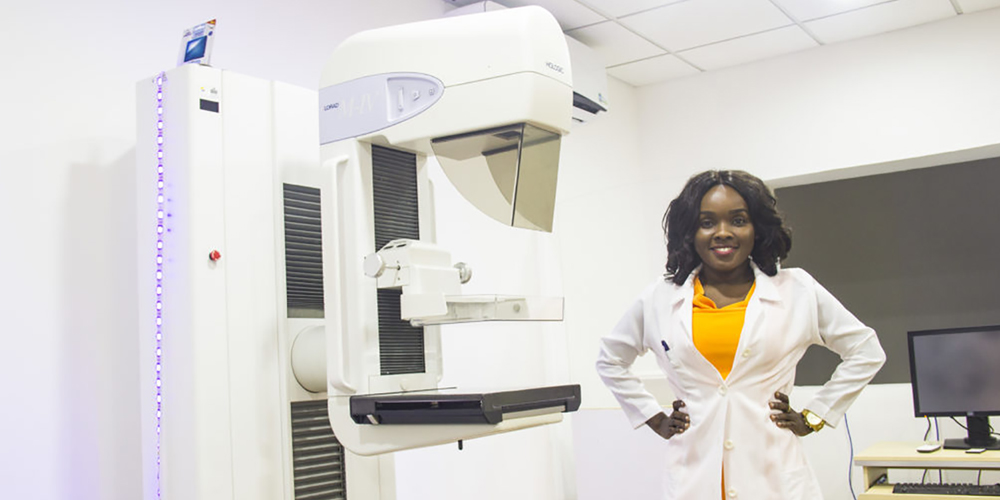
Digital Memmography
Digital mammography uses x-rays to easily detect abnormalities that otherwise may not be viewed clearly with traditional mammography. At Spectra Health, we are committed to finding the most invasive cancers as early as possible.
This is why we have the state-of-the-art Hologic M4 Platinum system to provide only the best service for our patients. We undertake both diagnostic and screening mammography for early detection and management of breast pathologies.
Our Advantages
Screening mammograms is performed within 10 to 12 minutes.
Allows for the radiologist to capture and manipulate images of the breast such that abnormalities are seen more easily.
Digital images are immediately available, easily stored and retrieved.
The digital machine is fast, so patients spend less time in uncomfortable positions.
Ultrasound
Ultrasound imaging is a safe, non-invasive technique which uses sound waves to produce pictures of the organs in the body. Spectra Heath offers a wide range of ultrasound services including general body examinations, doppler examinations for the imaging of vessels as well as obstetrics and gynaecological cases.
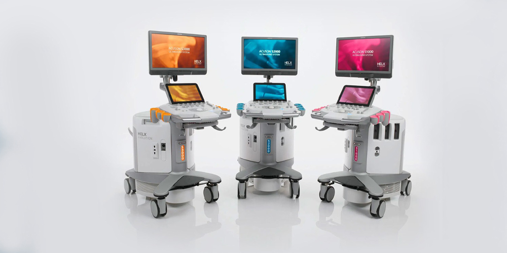
We also undertake ultrasound guided interventions. With an adjustable LCD flat panel ultrasound machine, our patients feel at ease and comfortable during every examination.
We also undertake ultrasound guided interventions. With an adjustable LCD flat panel ultrasound machine, our patients feel at ease and comfortable during every examination.
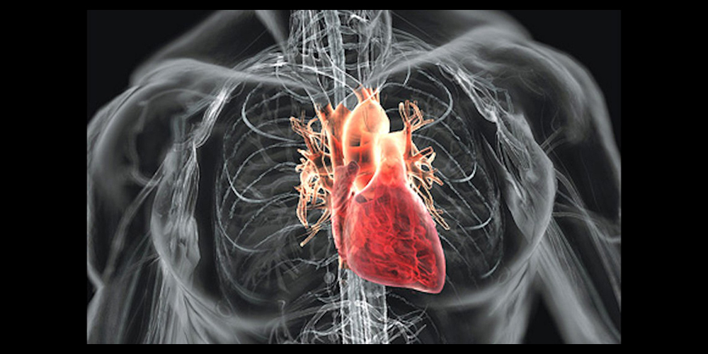
Echocardiography
Also referred to as echo test or heart ultrasound, and it takes “moving pictures” of the heart with sound waves. This test is employed to look at your heart’s structure and check how well your heart is working.
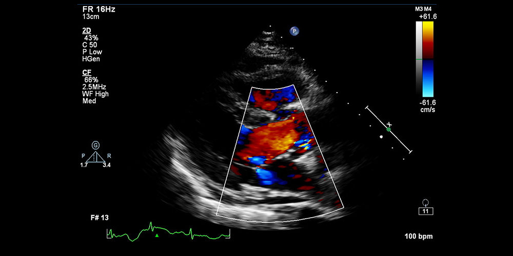
It is a safe procedure which helps to determine the health of a heart muscle especially after a heart attack.
Echo test is needed when ?
You have a heart murmur
You have unexplained chest pains
You’ve had rheumatic fever
You have congenital heart defect
The Test Can Show:
The size and shape of your heart
How well your heart is working overall
If a wall or section of your heart muscle is weak and not working correctly
If you have problems with your heart valve
If you have a blood clot
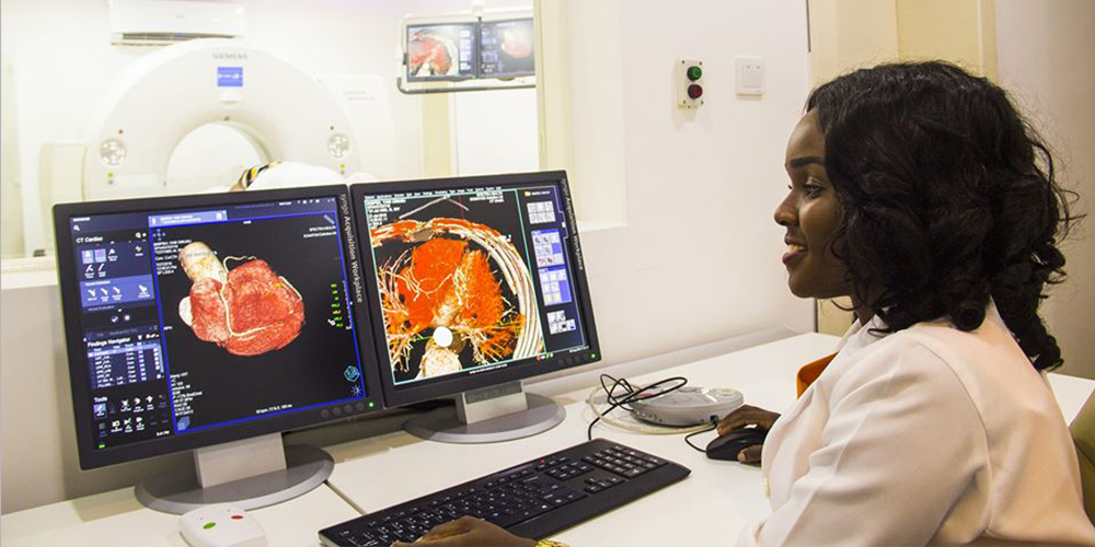
CT SCAN
A computerized tomography (CT) scan combines a series of X-ray images taken from different angles around your body and uses computer processing to create cross-sectional images (slices) of the bones, blood vessels and soft tissues inside your body. CT scan images provide more-detailed information than plain X-rays do.
A CT scan has many uses, but it is particularly well-suited to quickly examine people who may have internal injuries from car accidents or other types of trauma. A CT scan can be used to visualize nearly all parts of the body and is used to diagnose disease or injury as well as to plan medical, surgical or radiation treatment.
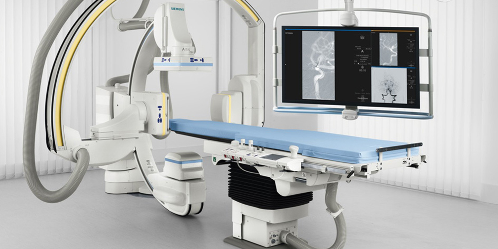
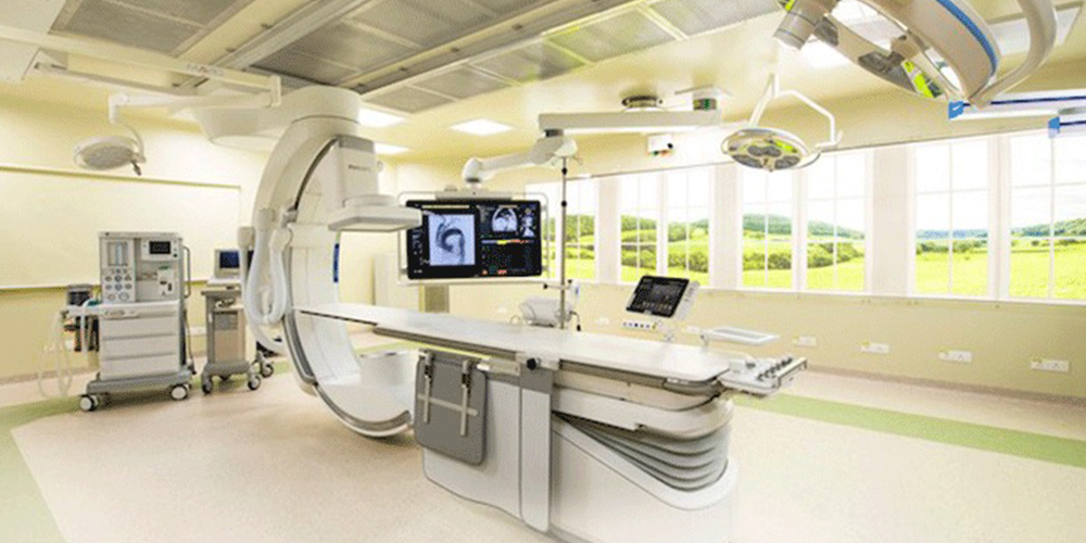
Angiography Suite
CATH lab: Cardiac catheterization is a procedure which requires that a thin, hollow tube (a catheter) be inserted into the blood vessel leading to the heart.
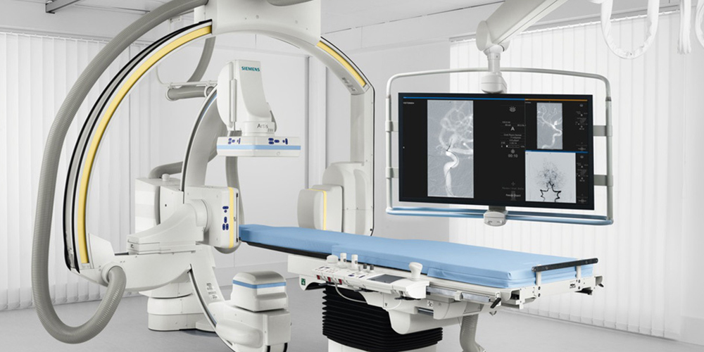
This is done to examine the heart and treat any stenosis or abnormalities found. This procedure is safe (complications may occur in 1 out of 200 cases) and can prevent a heart attack that could be life-threatening.
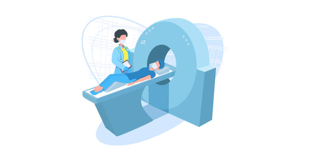
MRI Scanning
Magnetic resonance imaging (MRI) is a test that produces very clear pictures, or images, of the human body without the use of x-rays. MRI uses a large magnet, radio waves and a computer to produce these images. An MRI exam may provide further insight into an abnormality determined previously by an x-ray, ultrasound, or CT scan.
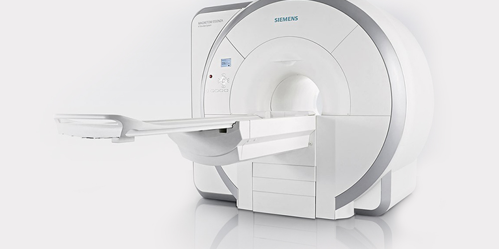
The advantage of MRI over past imaging techniques is that by
using magnetism instead of X-rays, MRI avoids exposing patients to ionizing radiation.
MRI is ideal for diagnosing and visualizing:
Multiple Sclerosis (MS)
Tumors of the pituitary gland and brain
Infections in the brain, spine or joints
Torn ligaments in the wrist, knee and ankle
Shoulder injuries
Tendonitis
Masses in the soft tissues of the body
Bone tumors, cysts and bulging or herniated discs in the spine
Strokes in their earliest stages
At Spectra Health, our MRI exams are performed using leading-edge MRI technology (Magnetom Essenza MRI (1.5T) and interpreted by experienced radiologists.
AppointmentOsteodensitometry
Osteodensitometry evaluates the strength of the skeleton by measuring the calcium content in the bones. This allows the analysis of the risk of fracture for the diagnosis and monitoring of osteoporosis (decrease in bone strength and increased risk of fracture).
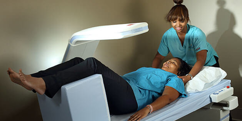
The purpose of a Bone Densitometry examination is the prevention of osteoporosis and, therefore, of fractures, particularly those of the hip and spine.
What to expect:
This examination is painless and without side effects. You lie on your back, motionless. Under the supervision of a technologist, the machine’s arm slowly passes over the body and emits low-dose x-rays and records the data to measure bone density.
Fluoroscopy
Fluoroscopy is a study of moving body structures – similar to an X-ray “movie.” A continuous X-ray beam is passed through the body part being examined, and is transmitted to a TV-like monitor so that the body part and its motion can be seen in detail.
Application:
Barium X-rays– fluoroscopy allows the physician to see the movement of the intestines as the barium moves through them
Cardiac catheterization– fluoroscopy enables the physician to see the flow of blood through the coronary arteries in order to evaluate the presence of arterial blockages.
Intravenous (IV) catheter insertion– fluoroscopy assists the physician in guiding the catheter into a specific location inside the body.
Interventional Cardiology
Interventional Cardiology is a subspecialty of cardiology in which catheter-based diagnostic tests and treatment are provided for coronary artery disease, valvular disease, structural heart disease, peripheral vascular disease and various other diseases.
In interventional cardiology, we avoid surgery through the use of tiny catheters, as with procedures like angioplasty and valve repair or replacement. We also perform diagnostic tests such as coronary angiograms, in which contrast or dye is placed in the coronary arteries and peripheral arteries to identify blockages in the vessels.
Our interventional cardiology team offers numerous treatment options for heart and vascular.
Conditions including:
Coronary artery disease (CAD)
Valvular heart disease
Structural and non-valvular heart disease
Hypertrophic cardiomyopathy (HCM)
Resistant hypertension
Atrial septal defect
Patent foramen ovale
Advantanges
The avoidance of the scars and pain, and long post-operative recovery.
Interventional cardiology procedure of primary angioplasty is now the gold standard of care for an acute myocardial infarction.
This involves the extraction of clots from occluded coronary arteries and deployment of stents and balloons through a small hole made in a major artery, which has given it the name “pin-hole surgery” (as opposed to “key-holesurgery”).
AppointmentDigital Platform
Digital experience monitoring (DEM) is a performance analysis discipline that
supports the optimization of the operational experience and behavior of a digital agent, human or machine, with the application and service portfolio of enterprises.
Schedule AppointmentAngiography
Angiography is a medical imaging technique used to visualize the inside, or lumen, of blood vessels and organs of the body, with particular interest in the arteries, veins and the heart chambers.
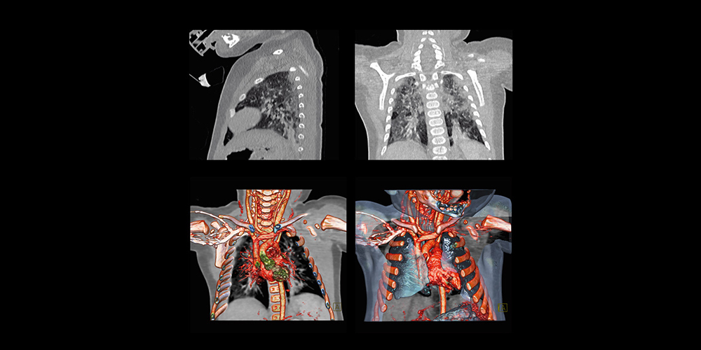
In coronary angiogram, our experts use CT scan to see your heart’s blood vessels. Coronary angiograms are part of a general group of procedures known as heart (cardiac) catheterization. The test is generally done to see if there’s a restriction in blood flow going to the heart and it is useful in diagnosing heart conditions such as atherosclerosis.
Process
During an angiogram, the iodine intravenous dye which is visible by the CT machine is injected into the blood vessels of your heart. The CT machine rapidly takes a series of images (angiograms), offering a look at your blood vessels. If clogged arteries are identified, angioplasty with stent placement may be performed.
Book Now
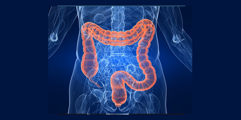
Colonoscopy
One of the early indications of possibly developing colon cancer is when abnormal growths in the colon known as polyps (usually not harmful) become cancerous.
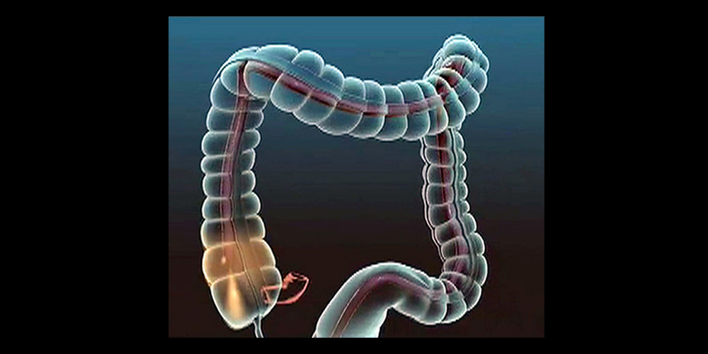
CT colonoscopy (sometimes referred to as virtual colonography) uses a low-dose CT scan to detect polyps and remove them when necessary. The procedure does not require sedation, and therefore safe and is much more comfortable than a conventional colonoscopy exam. Our state-of-the-art 64 slice Somatom Definition AS CT scan allows us to detect even minute abnormalities providing very clear and detailed images.
Process
A very small, flexible tube will be passed two inches into your rectum to allow air to be gently pumped into the colon. An electronic pump is used to deliver carbon dioxide gas into the colon. The purpose of the gas is to distend the colon as much as possible to eliminate any folds or wrinkles that might hide polyps from the physician’s view.
...
Next, the table will move through the scanner. Patients are asked to hold their breath for about 15 seconds or less before turning over and lying on their back or side for a second pass that is made through the scanner.
The entire examination is usually completed within 15 minutes.
Calcium Scoring
DCalcium scoring is a non-invasive outpatient test that can help determine the presence of coronary artery disease (CAD) and identify heart attack risk.
Very little preparation is needed, and testing typically takes less than 15 minutes without the need for an IV or medications.
Background
CAD (coronary artery disease or atherosclerosis) often starts as fatty substances that accumulate in the lining of one or more of the heart arteries. Over time, these deposits may become calcified (hardened with calcium). Angina (chest pain) can be caused by decreased blood flow through these hardened areas, restricting oxygen rich blood from reaching heart muscle. If these areas are not found and treated properly, the hardening could lead to a heart attack and other heart related problems.
Process
On the day of the test, a cardiac CT (scan of the heart) will capture detailed images of your heart and arteries. Using these images, our expert Radiologists can determine if you have any calcification (hardening) of the arteries, the location of these areas and the amount. All of this information will be used to determine your calcium “score”. With the help of this information, your physician can discuss your risks and develop a personalized plan of care, which may include dietary changes, the addition of other medications, or further testing.
Virtual Colonoscopy
Virtual colonoscopy or CT colonography is a procedure in which a radiologist uses x-rays and a computer to create images of your rectum and colon from outside the body. Virtual colonoscopy can show ulcers, polyps, and cancer (colorectal cancer).
Using a CT scanner and computer methods of reconstructing the images, the colon can be evaluated without a colonoscope and without sedation.
Process
Virtual colonoscopy takes the information produced by a CT scanner and processes this information to produce an image of the colon’s inner surface. When the colon is properly cleansed and expanded (distended)
...
with room air or carbon dioxide, and when the CT information is processed, our experienced radiologists can look at the inner lining or surface to detect polyps, ulcers or cancers. The procedure lasts about 10 to 15 minutes.
Screening Mammography
A screening mammogram is an x-ray exam of the breasts in a woman who has no symptoms. It is used to detect breast changes in women who have no signs or symptoms or new breast abnormalities. The goal is to detect cancer before clinical signs are noticeable.
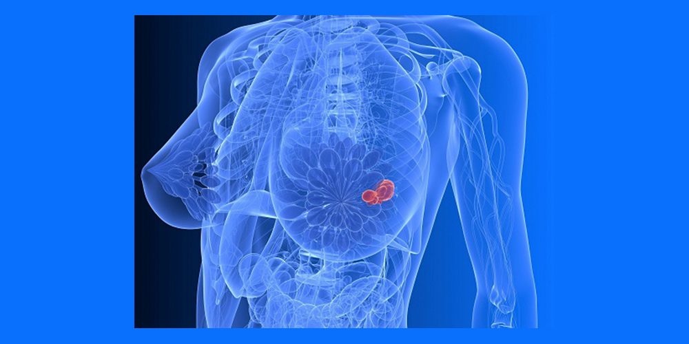
A screening mammogram is suggested annually for women age 40 and older
Process
During a mammogram, your breasts are compressed between two firm surfaces to spread out the breast tissue. Using our state-of-the-art Hologic
...
M4 Platinum system, we are able to capture and manipulate images of the breast such that abnormalities are seen more easily.
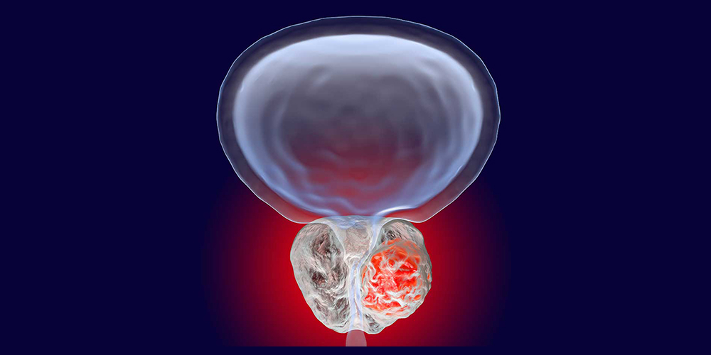
Prostate Cancer Screening
Prostate cancer is the most commonly occurring cancer in men. Whether or not a man should undergo screening for prostate cancer is an important decision.
Men at Greater than Average Risk
At age 40, men who are at increased risk of prostate cancer should include:
African American / Black men (Men of African Ancestry)
Men with a family history of prostate cancer in a first degree relative (father, brother, or son) diagnosed before age 65.
Process
African American / Black men (Men of African Ancestry)
A CT scan of the prostate reveals blood flow and anatomy of tissues in and around the prostate, allowing for the diagnosis and monitoring of tumor growth. A CT scan may be used to pinpoint the location of a tumor, evaluate the extent of cancer in the body and assess whether the disease is responding to treatment.
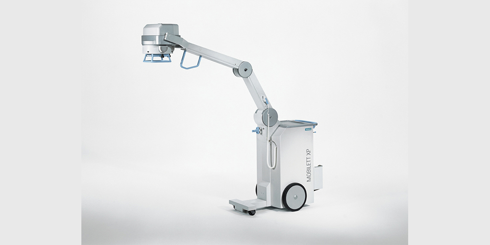
Digital X-Rays
X-rays are similar to light as they are a form of electromagnetic radiation, and an x-ray machine is similar to a camera. However, instead of using visible light, an x-ray machine uses x-rays to expose the image.
that supports the optimization of the operational experience and behavior of a digital agent, human or machine, with the application and service portfolio of enterprises....
Advantages
In the process of capturing a digital x-ray, digital image sensors are used instead of photographic film. This is the latest method of obtaining an x-ray and is preferred over the use of films as digital images are clearer and more accurate.
This advancement eliminates the need for chemical processing and longer wait times for film development as images are accessible for review immediately after your examination.
Images can also be sent to a physician electronically so he/she can access them on their desktop or personal computer and report their findings within minutes.
Process
During the examination, x-ray particles referred to as photons are sent by the x-ray machine to pass through the body, allowing a digital image to be recorded. This does involve exposure to low doses of x-rays to produce a black and white image on a computer screen. The amount of exposure is very small and directed towards the part of the body being imaged.
Laser Spine / Pain
Are you suffering from chronic pain in the neck and back? This condition is medically described as discogenic radiculopathy. Up until now, surgery had remained the only option for patients whose symptoms were not relieved by analgesics, physiotherapy and chiropractic sessions.

The good news is that Spectra Health now offers you resolution of your chronic neck and back pain without surgery!
Background of Discogenic Radiculopathy
A healthy spine has soft pliable pieces of disc material between the various vertebrae, which act as cushions and give the body flexibility. However, with aging, the discs undergo wear and tear and tend to lose their elasticity and strength. Aside the normal aging process, poor sitting, bending and lying posture are known to further accelerate the degenerative process. Protrusion and inflammation of the discs with attendant impingement on the surrounding nerves result in pain, back and neck stiffness, numbness and tingling sensation in the arms and legs.
Are you a candidate for Laser Spine care?
You have been diagnosed with spinal stenosis, bulging/herniated disc, sciatica or other chronic spine conditions.
You have already had open spine surgery and have not found relief.
You have not had success with conservative treatments like chiropractic care, physical therapy, yoga/stretching or prescription pain medication.
You have trouble sleeping, exercising, standing for long periods of time or participating in daily activities.
At Spectra Health, we pride ourselves by ensuring that our patients “don’t go another day with their chronic neck or back pain”
AppointmentDigital X-Rays
X-rays are similar to light as they are a form of electromagnetic radiation, and an x-ray machine is similar to a camera. However, instead of using visible light, an x-ray machine uses x-rays to expose the image.
that supports the optimization of the operational experience and behavior of a digital agent, human or machine, with the application and service portfolio of enterprises....
Advantages
In the process of capturing a digital x-ray, digital image sensors are used instead of photographic film. This is the latest method of obtaining an x-ray and is preferred over the use of films as digital images are clearer and more accurate.
This advancement eliminates the need for chemical processing and longer wait times for film development as images are accessible for review immediately after your examination.
Images can also be sent to a physician electronically so he/she can access them on their desktop or personal computer and report their findings within minutes.
Process
During the examination, x-ray particles referred to as photons are sent by the x-ray machine to pass through the body, allowing a digital image to be recorded. This does involve exposure to low doses of x-rays to produce a black and white image on a computer screen. The amount of exposure is very small and directed towards the part of the body being imaged.
Digital X-Rays
X-rays are similar to light as they are a form of electromagnetic radiation, and an x-ray machine is similar to a camera. However, instead of using visible light, an x-ray machine uses x-rays to expose the image.
that supports the optimization of the operational experience and behavior of a digital agent, human or machine, with the application and service portfolio of enterprises....
Advantages
In the process of capturing a digital x-ray, digital image sensors are used instead of photographic film. This is the latest method of obtaining an x-ray and is preferred over the use of films as digital images are clearer and more accurate.
This advancement eliminates the need for chemical processing and longer wait times for film development as images are accessible for review immediately after your examination.
Images can also be sent to a physician electronically so he/she can access them on their desktop or personal computer and report their findings within minutes.
Process
During the examination, x-ray particles referred to as photons are sent by the x-ray machine to pass through the body, allowing a digital image to be recorded. This does involve exposure to low doses of x-rays to produce a black and white image on a computer screen. The amount of exposure is very small and directed towards the part of the body being imaged.